3D Ultrasound Export: Volume, Cine, or Probe Record
Same symptom on your system?
Tell us your model and what it is doing. A GEPROBE engineer replies with a diagnosis path, part options and pricing — usually within 6 hours, with no obligation.
Chat on WhatsApp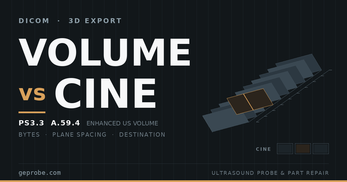
A 3D ultrasound export that reaches the archive as a spinning clip is rarely a dying transducer. It is usually a file that was never a volume, or a volume the receiving side cannot open.
Both possibilities are settled by numbers you can collect without opening a viewer: the size of the object, the spacing of its planes, and what the destination declared it supports.
This page works those three checks in that order, then turns the same evidence back onto the probe that produced the volume.
Size the Object Before Anyone Opens It
A volume has a floor size. An uncompressed frame occupies rows × columns × samples per pixel × bytes per sample — the same fixed length the standard uses to locate frames inside pixel data (PS3.3 C.7.6.3, 2026d, reviewed 2026-11-16).
Payload = frames × rows × columns × bytes per sample, where bytes per sample is Bits Allocated ÷ 8.
Take an invented case: 128 planes, 256 × 256 pixels, 16-bit samples.
That gives 128 × 256 × 256 × 2 = 16,777,216 bytes, about 16 MiB of pixel data before any header attribute. The dimensions are illustrative, not a claim about a named system.
Now compare that figure with what the archive actually holds. An instance of 1.4 MB filed under a 3D protocol, for an exam whose console reports 128 planes, is not the volume.
No viewer, no vendor call, no probe order.
| Archived bytes ÷ computed payload | Reading | Next move |
|---|---|---|
| 0.9 to 1.1 | Payload-sized, or compressed with a near-transparent syntax | Confirm the record class and the frame count, then stop |
| 0.3 to 0.9 | Compressed syntax, cropped region, or fewer frames than the console reported | Record the negotiated transfer syntax before deciding |
| Below 0.1 | Not the volume: a rendered clip, a still, a thumbnail | Read the record class; leave the probe alone |
| Above 1.1 | Payload plus derived objects, or a mistyped dimension in your arithmetic | Re-read the four numbers from the header |
The four numbers sit in the header: frames, rows, columns, bits allocated.
Write them beside the record class name. A volumetric object is filed as Enhanced US Volume Storage.
Derived and screen-captured data arrives as Ultrasound Multi-frame Image, Ultrasound Image or Secondary Capture (PS3.4 Table B.5-1, reviewed 2026-11-16).
One caveat travels with any volumetric record. Conformance proves how data is encoded, not how faithful the scan conversion was.
Building the Cartesian volume, its relation to the source frames, and the rendering algorithms all sit outside the standard (PS3.17 PP.3, reviewed 2026-11-16). A clean object can still hold a badly reconstructed volume.
The Volume Is Also a Probe Record
A genuine volume is not only an archive object. It is the most complete probe diagnosis record the system produces.
Every plane in it was placed by the sweep that produced it, and the standard requires neighbouring planes to sit at even spacing along the volume's Z axis (PS3.3 A.59.4.1.2, reviewed 2026-11-16).
So measure the spacing rather than trusting the picture. Read each plane's position, difference consecutive positions, then look at the spread of those differences.
Δz_k = z_(k+1) − z_k for k = 1 … n − 1. CV = standard deviation of Δz ÷ mean Δz.
CV is the uniformity figure for one sweep. Put the limit in your own procedure; a workable house band is 0.02 to pass and 0.05 to re-run before escalating. That band is our suggestion, not a standard.
| What the volume shows | Where it usually originates | First test |
|---|---|---|
| A stripe holding the same azimuth position in every plane | One element, its channel path, or a connector pin | Re-seat the connector, then read the system's element test |
| Whole planes missing, or a gap mid-sweep | Frames dropped in transfer, or a stalled sweep | One re-export, then a repeat sweep at a second angle |
| Spacing that fans out towards the end of the sweep | Motor or encoder linearity | Two sweeps at two sweep speeds |
| A visible seam in a 360° acquisition | Position calibration, not the array | Re-run with the vendor's calibration artefact |
Rows one and two are the ones that reach a repair bench, and they point in opposite directions.
A stripe that holds its azimuth position and follows the probe across hosts is array or cable work. A missing block of planes deserves one re-export before anyone calls it a transducer.
Where the finding does sit in the probe, the work is component level.
That covers element-array restoration, connector pin repair, EEPROM reprogramming so the host identifies the transducer again, and resealing after fluid ingress (GEPROBE probe repair, reviewed 2026-11-16).
Pair the Transducer With the Destination Before You Buy
A volume-capable transducer is bought for what it renders on the cart. Whether that output survives the network depends on four questions that have nothing to do with the probe.
- Will anyone need to re-slice the volume off-axis after the patient has left the room?
- Does the department hold a review workstation that opens this vendor's volume format?
- Does the target archive accept a volumetric ultrasound record, or only multi-frame and single-image ultrasound records?
- Is the volume itself the record of the exam, or does a rendered clip satisfy the documentation requirement?
Answer one and three together first. If re-slicing is required and the archive takes no volumetric record, the gap is on the receiving side. A newer transducer does not close it, and that budget belongs to integration work.
If re-slicing is not required and a clip satisfies documentation, the probe still earns its price. Live rendering, on-cart measurement and immediate clinical feedback all stay local.
Plan the archive around cine in that case, and stop treating the absent volume as a defect.
The expensive middle case is the one to avoid. Buying the transducer for offline MPR that nobody has yet confirmed the archive can deliver produces a capable probe and an unchanged workflow.
When the Archive Cannot Hold the Volume
If no DICOM volume can land, the console becomes the only copy. That is a records problem, and it surfaces at decommission rather than at acquisition.
Three habits keep it manageable. Export volumes to approved media on a schedule you can name.
Keep one review workstation as the designated reader, with its software revision written down beside it. Record where each exam's volume really sits, so a later re-send request has an address.
At resale or decommission, be explicit about which of two assets is moving.
The first is capture capability on the cart. The second is an archive copy the buyer can open. They are different purchases, and a buyer who assumes the second when only the first exists has no recourse once the cart leaves the building.
Return-to-Service Gates After a Volume Probe or Drive Repair
A repaired probe that renders nicely on the cart has not been accepted. Seven gates close the loop, and the last two are the ones teams skip.
| Gate | Pass condition | Evidence to file |
|---|---|---|
| Console sweep | A, B and C planes rebuild and reslice interactively | Dated screenshot set |
| Frame count | Inside the pre-repair count for the same preset | Before and after numbers |
| Plane spacing | CV inside your house limit on three consecutive sweeps | Δz list and the CV |
| Image quality | 2D, 3D and 4D modes restored, no repeating stripe | Paired images, identical preset |
| System recognition | Host names the correct model, no degraded mode | Service screen capture |
| Export re-test | One synthetic volume reaches the usual destination as the same record class | Class name, byte size, destination |
| Paperwork | Test report and cosmetic checklist attached to the work order | Document identifier |
The export re-test catches a repair that fixed the probe and left the destination unable to receive what the probe now produces.
It also catches the opposite case, where the probe always worked and only the export path was ever broken.
Run it with a phantom, never with patient data, and keep the files until the work order closes.
What GEPROBE Tests and What Belongs Elsewhere
GEPROBE repairs probes and boards. It does not configure PACS destinations or write DICOM integrations, and saying so is more useful than pretending otherwise.
On the probe side the published scope is free evaluation with a written failure report, OEM-standard restoration in a cleanroom, and imaging verification on original host systems before return.
A test report with a cosmetic checklist comes with the work (GEPROBE repair, reviewed 2026-11-16). Console-side replacements are catalogued separately (ultrasound parts, reviewed 2026-11-16).
Price and terms follow the evaluation of the actual unit. Nothing on this page should be read as a lead-time commitment.
A useful enquiry carries four things. Give the system model and software revision, and the probe or board part number with label photos taken by qualified staff.
Say whether 3D or 4D fails on the cart or only after export. Add quantity and destination country so shipping can be answered in the same reply.
Sources
- DICOM PS3.3 C.7.6.3, image pixel attributes — reviewed 2026-11-16.
- DICOM PS3.3 A.59.4, Enhanced US Volume functional groups — reviewed 2026-11-16.
- DICOM PS3.4 B.5, standard SOP classes — reviewed 2026-11-16.
- DICOM PS3.17 PP.3, 3D ultrasound solutions — reviewed 2026-11-16.
- GEPROBE probe repair — reviewed 2026-11-16.
- GEPROBE ultrasound parts — reviewed 2026-11-16.
Related Articles
Ultrasound QA Baseline Drift: Triage Before You Repair
Ultrasound QA baseline drift, worked in order: a measured noise floor, a one-session swap design, a cost threshold, and what a clean image cannot prove.
Qualifying a Refurbished Ultrasound Parts Supplier
Sample size, landed cost, response clocks and a repair ceiling: the arithmetic to run before a refurbished ultrasound parts trial order.
Used Ultrasound Export UK: Prove Equipment, Not Waste
How to prove a used ultrasound is equipment rather than waste on a used ultrasound export UK shipment, with the probe-level evidence most files miss.